

Retinal Detachment Treatment at Thakur Eye Hospital
Advanced Retina Care for Permanent Vision Protection
Retinal Detachment is a serious and sight-threatening eye condition that requires immediate medical attention. It occurs when the retina separates from its normal position at the back of the eye.
The retina is a thin, light-sensitive layer of tissue that plays a vital role in vision by converting light into signals that are sent to the brain. When the retina detaches, its blood supply is disrupted, and if not treated promptly, it can lead to irreversible vision loss or blindness.
At Thakur Eye Hospital, we understand the urgency and emotional stress associated with retinal detachment. Our experienced retina specialists provide comprehensive diagnostic evaluation, advanced surgical care, and personalized post-treatment follow-up to help restore retinal attachment and preserve vision. With cutting-edge technology and a patient-centric approach, we strive to deliver the highest standards of retinal care.
Book a Consultation today to protect your vision and receive expert guidance from our retina specialists.
What Is Retinal Detachment?
Retinal detachment occurs when the retina separates from the underlying tissue that supports it. This can interfere with retinal function and, if untreated, may cause permanent vision loss.
Retinal detachment can develop suddenly and is often painless, so warning symptoms may be overlooked. It is a medical emergency that requires prompt ophthalmic assessment. The anatomical and visual outcome depends on factors such as the type, location, extent and duration of the detachment and whether the central retina is involved. Early diagnosis and timely treatment are therefore important.
Causes & Risk Factors of Retinal Detachment
Retinal detachment can occur for different underlying reasons. Identifying the type of detachment, associated retinal tears or traction, and relevant risk factors helps the retina specialist determine an appropriate treatment approach.
Risk factors can include age-related vitreous changes, retinal tears or holes, eye trauma, high myopia, previous eye surgery, diabetic retinopathy, inflammation or infection, and a family history of retinal detachment. People at increased risk should seek prompt assessment if they develop new flashes, floaters, a curtain-like shadow or sudden loss of vision.
Recognizing Symptoms (When to Seek Immediate Help)
The symptoms of retinal detachment often appear suddenly and should never be ignored. Early recognition can be vision-saving.
Common warning signs include:
- Sudden appearance or increase in floaters (black dots, lines, or cobweb-like shapes)
- Flashes of light, especially in peripheral vision
- A dark shadow or curtain moving across part of the visual field
- Blurred, distorted, or reduced vision
- Sudden loss of side (peripheral) vision
If you experience any of these symptoms, seek emergency eye care immediately. Delaying treatment may significantly reduce the chances of visual recovery.
How Retinal Detachment Is Diagnosed
Accurate diagnosis is the foundation of effective treatment. At Thakur Eye Hospital, we use advanced diagnostic technology to evaluate the retina in detail and determine the extent of detachment.
Diagnostic methods include:
- Dilated fundus examination to directly visualize the retina
- Optical Coherence Tomography (OCT) for high-resolution cross-sectional images
- B-scan ultrasound imaging when visibility is limited due to bleeding or cataracts
- Visual acuity and visual field testing to assess functional vision loss
These investigations help our retina specialists create a customized treatment plan based on the severity, location, and cause of detachment.
Available Treatments for Retinal Detachment
The treatment plan for retinal detachment depends on the type and location of the detachment, the retinal tear or traction involved, the condition of the macula and the overall health of the eye.
Laser photocoagulation and cryotherapy: These may be used for selected retinal tears or limited detachments. They create an adhesion around the retinal break to reduce the risk of fluid passing through it.
Pneumatic retinopexy: In selected uncomplicated detachments, a gas bubble may be placed inside the eye to support the retina while laser or cryotherapy seals the retinal break. Postoperative head positioning may be required.
Scleral buckling: A silicone buckle is placed on the outside of the eye to reduce traction and support retinal reattachment. It is an established technique for selected types of retinal detachment.
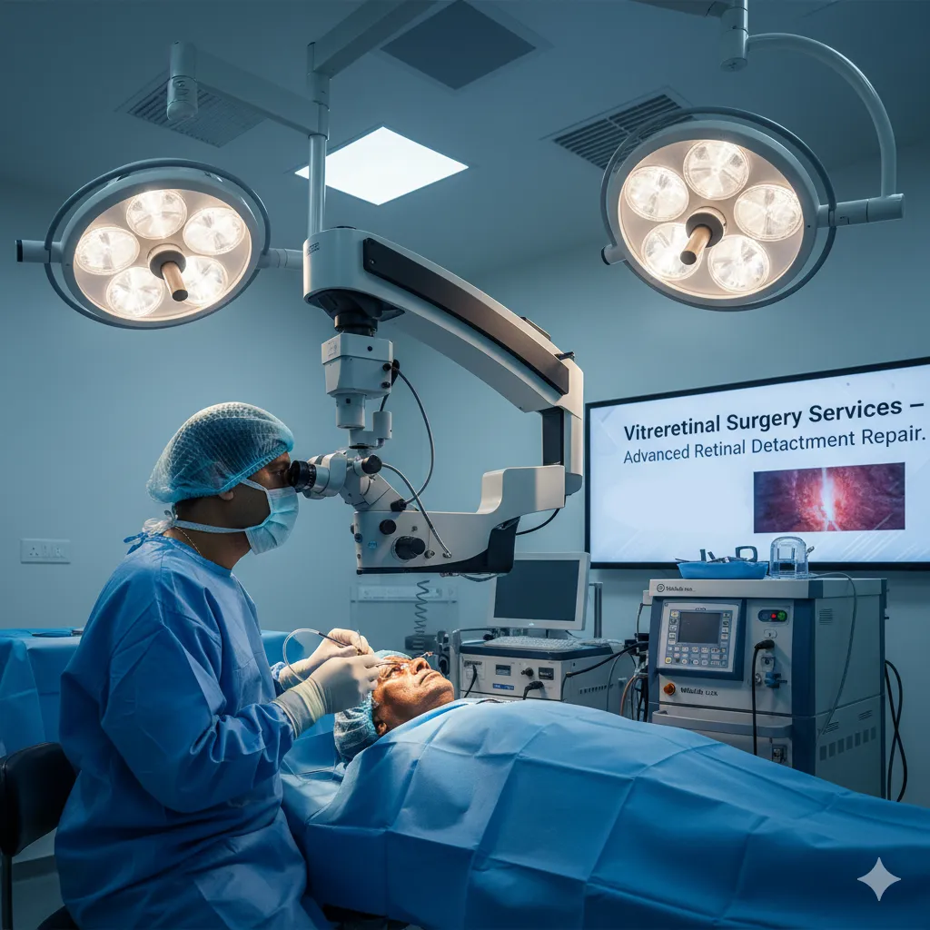
Vitrectomy: Vitrectomy is commonly used for selected complex detachments, including cases involving vitreous traction, scar tissue, diabetic retinopathy or trauma. Gas or silicone oil may be used to support the retina after internal repair.
At Thakur Eye Hospital, retinal-detachment treatment is planned by experienced retina surgeons according to the individual retinal findings. Anatomical and visual outcomes vary with the severity and characteristics of the detachment.
Preparing for Retinal Detachment Surgery
Proper preparation plays a vital role in surgical success and smooth recovery.
Before surgery, patients typically undergo:
- Detailed consultation and retinal imaging
- Review of medical history and medications
- Instructions regarding fasting, if required
- Guidance on temporary medication adjustments
- Arrangements for transportation and post-surgery care
Our team ensures that patients are well-informed and comfortable before proceeding with treatment.
Recovery & Post-Operative Care
Recovery after retinal detachment surgery varies depending on the procedure performed and the severity of the condition.
Post-operative care usually includes:
- Protective eye shields and prescribed medications
- Avoiding heavy lifting, bending, and strenuous activity
- Maintaining specific head positioning if advised
- Attending scheduled follow-up appointments
- Monitoring gradual vision improvement over weeks to months
While some visual recovery may be noticed early, complete healing and stabilization can take several months. Regular follow-up is essential for long-term success.
Long-Term Outlook After Retinal Detachment Treatment
Visual outcomes depend on:
- How early the detachment was treated
- Whether the macula (central vision area) was involved
- The underlying cause of detachment
Early intervention often results in better vision preservation. Even in complex cases, surgery can prevent further vision loss and significantly improve quality of life.
Why Choose Thakur Eye Hospital for Retinal Detachment Treatment?
Patients trust Thakur Eye Hospital for retinal care because of our:
- Experienced retina specialists
- Advanced diagnostic and surgical technology
- Comprehensive treatment options under one roof
- Patient-first and transparent approach
- Dedicated post-operative care and follow-up
- Strong focus on safety, comfort, and outcomes
Your vision is our priority, and we are committed to delivering care that meets the highest medical standards.
FAQs, Retinal Detachment
Is retinal detachment surgery urgent?
Yes. Retinal detachment is a medical emergency, and early treatment is critical to prevent permanent vision loss.
Can retinal detachment recur?
Yes, recurrence is possible. Regular follow-ups and timely treatment reduce this risk.
Will vision fully recover after surgery?
Visual recovery varies according to the type, location and duration of the detachment and the condition of the retina. Prompt assessment and treatment are important because delays can reduce the potential for visual recovery.
Is retinal detachment surgery painful?
No. Surgery is performed under anesthesia, and post-operative discomfort is managed with medication.
How soon can normal activities be resumed?
Light activities may resume within days, but complete recovery depends on the procedure and individual healing.